
The Heart Attack: What It Is, What It Feels Like, and What to Do
Heart attacks are not always dramatic or obvious. Learn how to recognise possible symptoms, why fast action matters, and what workplaces can do to respond.
A complete guide to myocardial infarction, from ancient Egypt to the modern emergency department
Need the short version?
This is the full guide to what a heart attack is, how it develops, and why it affects different people differently. If you need to know what to do right now, we have a shorter, action-focused companion post: Heart Attack: What to Do.
Every year, around 100,000 people in the UK are admitted to hospital following a heart attack.[1] That is one admission roughly every five minutes. Despite decades of medical progress, heart disease remains the leading cause of death worldwide, killing an estimated 17.9 million people annually.[2] Yet most people could not accurately describe what a heart attack is, what triggers one, or what to do in the critical minutes before an ambulance arrives.
This post sets out to change that. It covers the history of how humanity came to understand the heart attack, the biology of what actually happens inside the body, how to recognise one in yourself or someone else (including the presentations that are frequently missed), and what you can do to help. Because in a cardiac emergency, bystanders are not a backup option: they are often the only option.
A Condition as Old as Civilisation
Ancient Recognition
The heart attack is not a modern affliction. Evidence of coronary artery disease has been found in Egyptian mummies dating to around 1550 BCE.[3] The Ebers Papyrus, one of the oldest surviving medical documents, describes a condition in which "the heart is overpowered" and the left arm feels weak, a description that maps strikingly well to what we now recognise as cardiac pain. Ancient Egyptians attributed such symptoms to the heart being the seat of emotion and spirit; treatment was accordingly ritualistic rather than physiological.
Greek and Roman physicians including Hippocrates and Galen recognised the heart as central to life, though their understanding of its mechanics was incomplete. Galen believed the heart produced a vital spirit rather than functioning as a pump. The idea that blood circulated, rather than being consumed and replenished, would not be demonstrated for another 1,400 years.
The Circulation Revolution
In 1628, the English physician William Harvey published [4]De Motu Cordis, demonstrating for the first time that the heart functions as a pump and that blood circulates continuously around the body. This was a foundational moment: without understanding circulation, the concept of a blocked coronary artery causing localised heart muscle death was simply not available to medicine.
Over the following two centuries, anatomists began identifying the coronary arteries and describing what happened when they became diseased. In 1768, the English physician William Heberden gave the first formal clinical description of angina pectoris, the chest pain caused by insufficient blood supply to the heart.[5] He described patients who, on walking uphill, experienced a "strangling and anxious" sensation in the chest that forced them to stop. He noted, with characteristic understatement, that the condition "seems not to threaten immediate danger." Sometimes it did not. Often it did.
Naming the Event
The specific event we now call a heart attack, where a coronary artery becomes completely blocked and a section of heart muscle dies, was not clearly described until the early twentieth century. In 1912, the American cardiologist James Herrick published a landmark paper identifying the clinical features of coronary artery occlusion and arguing that patients could survive such an event.[6] This was a genuinely radical claim: the prevailing view was that complete coronary occlusion was invariably fatal. Herrick described patients who had survived, connecting their symptoms to the pathological findings observed at post-mortem in those who had not.
The term "myocardial infarction" (MI) derives from the Latin [7]infarctus, meaning stuffed or obstructed, and myocardium, the muscular tissue of the heart. The infarct is the area of tissue that has died due to loss of blood supply. The colloquial term "heart attack" came into widespread use in the mid-twentieth century as the condition became a major public health concern.
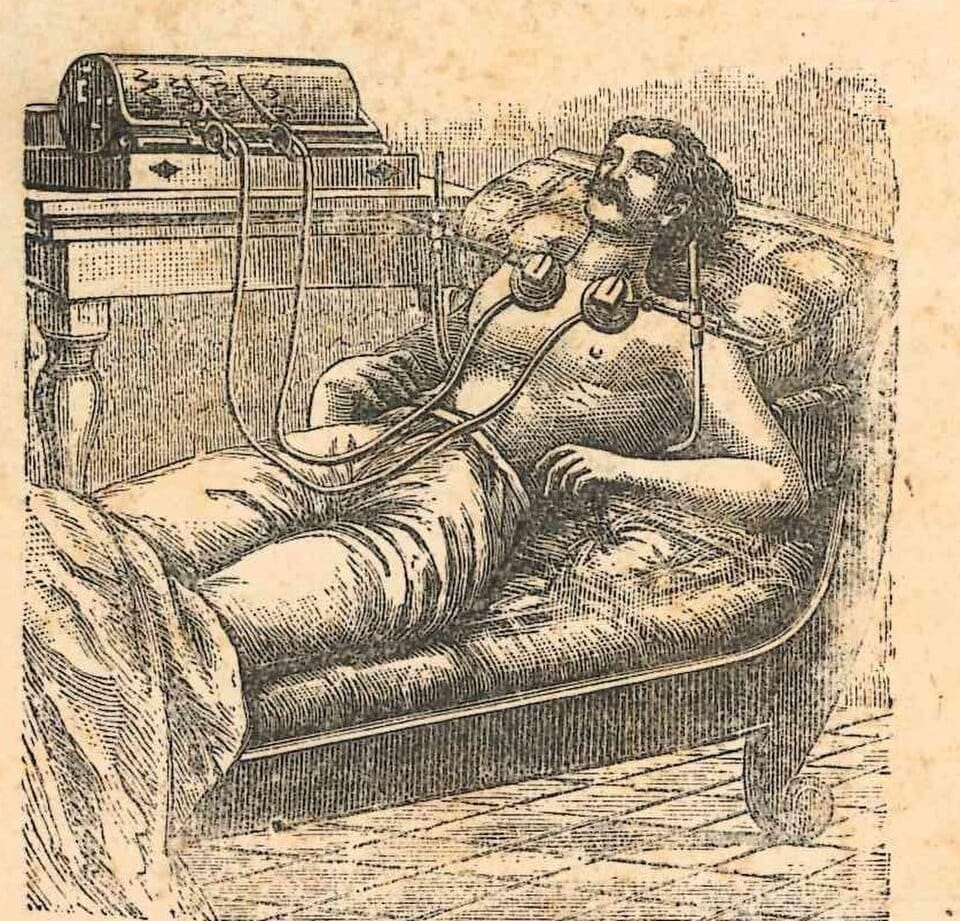
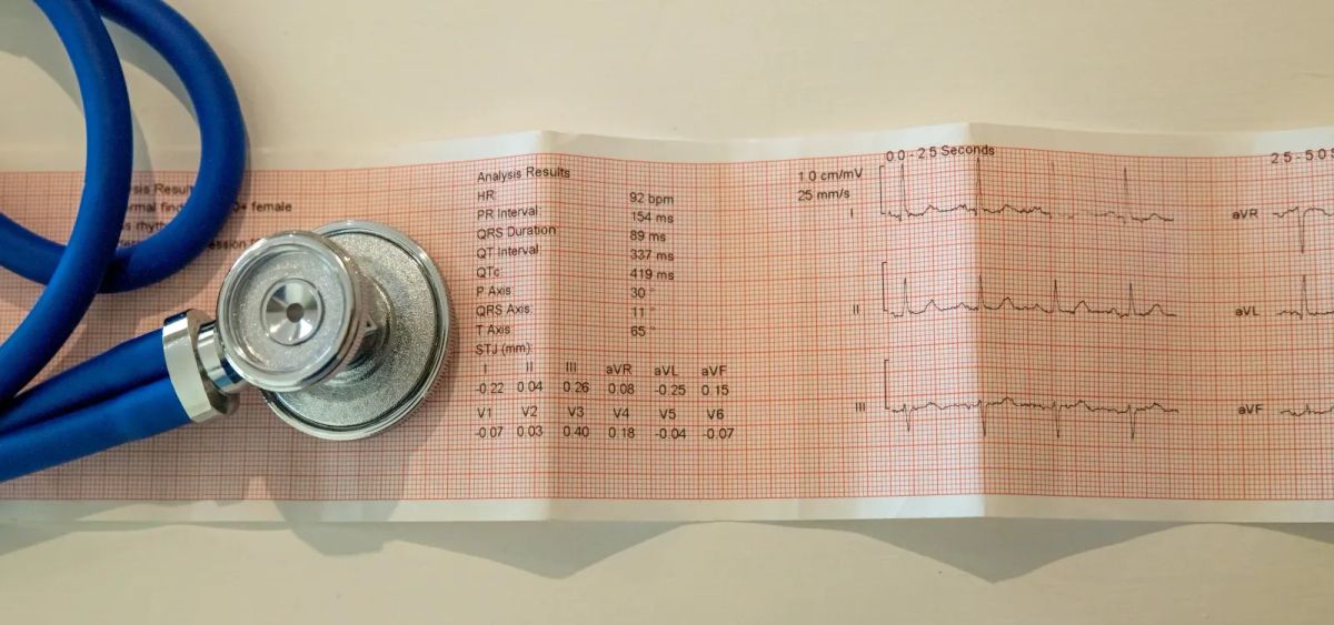
The Electrocardiogram and the Twentieth Century
The electrocardiogram (ECG) transformed cardiac diagnosis. Developed by the Dutch physiologist Willem Einthoven in the early 1900s (for which he received the Nobel Prize in 1924), the ECG allowed clinicians to detect the electrical changes in the heart caused by muscle damage.[8] This gave medicine a tool for diagnosing heart attacks in living patients rather than only identifying them at post-mortem.
Through the mid-twentieth century, treatment remained largely passive: bed rest for weeks, sometimes months. The logic was that the damaged heart needed complete rest to heal. This approach was later shown to be harmful in many cases, but it dominated until the 1970s.
The development of coronary care units (CCUs) in the 1960s marked a turning point. For the first time, patients were monitored continuously and could be treated immediately if they developed dangerous arrhythmias, the abnormal heart rhythms that are a common and life-threatening complication of MI. Defibrillation, which had been demonstrated experimentally in the 1950s, became a practical emergency intervention.[9]
Thrombolytics, Stents, and Modern Intervention
The 1980s brought the understanding that most heart attacks are caused by a blood clot (thrombus) forming on a ruptured atherosclerotic plaque in a coronary artery, and that dissolving or removing the clot could restore blood flow and save heart muscle. Thrombolytic ("clot-busting") drugs such as streptokinase and, later, tissue plasminogen activator (tPA) became standard treatments.[10]
Percutaneous coronary intervention (PCI), in which a catheter is threaded through the arteries to the site of the blockage, a balloon inflated to open the vessel, and a stent inserted to keep it open, progressively replaced thrombolytics as the gold-standard treatment for the most serious type of heart attack. The speed with which PCI is performed is directly related to outcomes: every minute of continued blockage means more muscle death. "Door to balloon" time (from hospital arrival to vessel opening) is now a closely monitored quality metric.[11]
Today, a person who reaches a specialist cardiac centre quickly and receives prompt PCI has a survival rate that would have seemed remarkable to the physicians of even fifty years ago. The challenge that remains is the gap between symptom onset and calling for help, a gap that first aid awareness directly addresses.
What Actually Happens: The Biology of a Heart Attack
The Coronary Arteries
The heart is a muscle. Like all muscles, it requires a continuous supply of oxygenated blood to function. That supply comes through the coronary arteries: two main vessels that branch from the aorta just above the heart and spread across its surface, delivering blood directly to the myocardium (heart muscle).
The left coronary artery divides into the left anterior descending (LAD) artery, supplying the front and main pumping chamber of the heart, and the circumflex artery, supplying the left side. The right coronary artery (RCA) supplies the right side and the inferior portion of the heart. Blockages in different vessels produce different patterns of damage, which is why the location of a heart attack matters clinically.
Atherosclerosis: The Slow Build
Most heart attacks do not happen suddenly out of nowhere. They are the endpoint of a process that typically develops over decades. That process is atherosclerosis: the gradual accumulation of fatty deposits, inflammatory cells, and fibrous tissue within the walls of the coronary arteries.[12]
The process begins when the inner lining of an artery (the endothelium) is damaged, often by high blood pressure, smoking, elevated blood glucose, or the physical shear stress of turbulent blood flow. Low-density lipoprotein (LDL) cholesterol penetrates the damaged lining and becomes oxidised. This triggers an immune response: white blood cells migrate into the arterial wall, engulf the oxidised LDL, and become foam cells. Over time, a plaque forms: a raised deposit of fatty material, inflammatory cells, and fibrous tissue within the arterial wall.
Plaques narrow the artery lumen (the space through which blood flows), which can cause angina, the chest pain on exertion that occurs because the heart cannot get enough blood during increased demand. But many plaques that trigger heart attacks are not the largest ones. Smaller, "vulnerable" plaques with a thin fibrous cap overlying a large lipid core are prone to rupture.
Plaque Rupture and Thrombosis
When a vulnerable plaque ruptures, the lipid core is exposed to circulating blood. The body responds as it does to any wound: platelets rush to the site and begin forming a clot. In a coronary artery, this clot can grow rapidly, partially or completely blocking blood flow to the downstream heart muscle.[13]
A partial blockage may cause unstable angina or a non-ST-elevation MI (NSTEMI), serious but with preserved blood flow to some degree. A complete blockage causes an ST-elevation MI (STEMI): the classic heart attack in which a full-thickness section of heart muscle is at immediate risk. The STEMI requires the fastest possible intervention.
The Ischaemic Cascade
Once blood supply is cut off, the affected muscle cells begin to die in a process called ischaemic cascade. Within seconds, affected cells switch from aerobic to anaerobic metabolism. Within minutes, cellular function begins to deteriorate. After around 20 minutes of complete ischaemia, irreversible cell death begins.[14] After six hours without reperfusion, the damage is typically extensive and permanent.
This is why time is the central variable in heart attack outcomes. The phrase used in emergency cardiology is "time is muscle": every minute of continued ischaemia means more myocardial cells are lost. Lost heart muscle does not regenerate. The function that goes is gone.
Key fact: time is muscle
Irreversible heart muscle death begins within approximately 20 minutes of a coronary artery becoming completely blocked. After six hours, damage is typically extensive. The fastest possible recognition and emergency response directly determines how much heart muscle survives.
Recognising a Heart Attack
The Classic Presentation
The textbook heart attack presents with central chest pain, typically described as a crushing, squeezing, pressing, or heavy sensation. Patients often describe it as feeling like an elephant sitting on the chest, or a tight band around the thorax. The pain may radiate to the left arm, jaw, neck, or back.[15]
Accompanying symptoms commonly include breathlessness, sweating (often described as a cold or clammy sweat), nausea or vomiting, dizziness or light-headedness, and a profound sense of impending doom, an intense anxiety that something is badly wrong. The person may appear pale or grey.
These symptoms may develop suddenly or build over minutes. They do not reliably go away with rest (unlike angina, which typically eases within a few minutes of stopping activity). Symptoms lasting more than 15 minutes that do not resolve should be treated as a medical emergency.
What People Often Dismiss
A significant proportion of people delay calling for help because their symptoms do not match the dramatic portrayal of a heart attack in popular culture. Common reasons for delay include attributing symptoms to indigestion, muscle pain, or anxiety; hoping the discomfort will pass; and reluctance to cause a fuss or be wrong.[16]
The average delay between symptom onset and calling emergency services in the UK is around two to three hours.[17] That delay costs lives. In the time it takes most people to decide the symptoms are serious enough to act on, irreversible myocardial damage is already underway.
When in doubt, call 999
If you or someone else has chest pain or other symptoms of a heart attack that have lasted more than a few minutes, call 999 immediately. Do not drive yourself. Do not wait to see if it gets better. The cost of being wrong is embarrassment. The cost of waiting is potentially fatal.
Atypical Presentations: Who Gets Missed and Why
The classic chest-clutching, dramatic collapse that most people associate with a heart attack is one presentation. It is not the only one, and it is not even the most common in some groups. Understanding atypical presentations is not a minor footnote — it is a matter of life and death for a substantial proportion of heart attack patients.
Heart Attacks in Women
Women are significantly more likely than men to present with atypical symptoms, and significantly more likely to be misdiagnosed or to have the seriousness of their condition underestimated.[18] Research part-funded by the British Heart Foundation found that women had a 50% higher chance than men of receiving an incorrect initial diagnosis following a heart attack.[19]
While chest pain remains common in women, it is less likely to be the dominant or presenting symptom. Women are more likely to experience:
- Shortness of breath without chest pain
- Nausea, vomiting, or indigestion-like discomfort
- Back pain or jaw pain rather than left arm radiation
- Unusual fatigue, sometimes for days before the event
- Pain in the upper abdomen
- Dizziness or light-headedness
- Sleep disturbance in the days preceding the event
Some of these symptoms, taken individually, are easy to attribute to other causes. Fatigue is common. Nausea is common. Back pain is common. It is the combination, the context, and the clinical judgement applied to them that matters, and that judgement has historically been applied less accurately to women than to men.
Part of the explanation is biological. Women more frequently experience a type of coronary artery disease called spontaneous coronary artery dissection (SCAD), which tends to occur in younger women and produces different ECG patterns.[20] Women are also more likely to have microvascular disease, affecting the very small coronary vessels rather than the major arteries, which may not show up on standard angiography.
Another part is systemic. Heart disease has historically been researched, modelled, and clinically understood primarily through male populations. The consequences of that research gap continue to affect diagnostic accuracy.
Awareness matters here not just for clinicians but for individuals. Women having a heart attack often do not recognise it as such, and therefore delay seeking help. A woman who wakes at 3am with unusual fatigue, nausea, and a vague sense that something is wrong should not dismiss it. She should call 999.
Atypical Presentations in South Asian Populations
People of South Asian origin (broadly: those with family origins in India, Pakistan, Bangladesh, Sri Lanka, and Nepal) have a significantly higher rate of coronary artery disease than the general UK population, and tend to develop it at a younger age.[21] The risk is estimated at approximately 50% higher than in white European populations. The reasons are multifactorial and include genetic predisposition, higher rates of type 2 diabetes and insulin resistance, different patterns of fat distribution (with more visceral fat at a given BMI), and lower average HDL ("good") cholesterol.
South Asian patients are more likely to present with diabetes as a comorbidity, and diabetes substantially alters the pain experience of a heart attack. Diabetic autonomic neuropathy can blunt or abolish the chest pain response, leading to "silent" or near-silent myocardial infarctions that are only detected retrospectively through ECG changes or cardiac enzyme tests.[22]
The practical implication is that a South Asian individual, particularly one with diabetes, who feels unusual fatigue, breathlessness, or a vague chest discomfort, even without dramatic chest pain, warrants immediate medical assessment.
Atypical Presentations in Black African and Caribbean Populations
Black African and Caribbean populations in the UK have lower rates of coronary artery disease than South Asian or white European populations, but significantly higher rates of hypertension (high blood pressure) and haemorrhagic stroke.[23] When heart attacks do occur, they are more frequently associated with hypertensive heart disease, where chronic high blood pressure causes structural changes to the heart muscle (hypertrophy) that alter the clinical picture.
As with South Asian populations, higher rates of type 2 diabetes in some Black communities create a similar risk of atypical or silent presentation. There are also well-documented disparities in the speed and quality of care received by Black patients presenting to emergency departments, an issue of healthcare access and systemic bias that affects outcomes independent of the underlying disease.[24]
Silent Heart Attacks
A silent myocardial infarction (SMI) is one that produces few or no recognisable symptoms at the time it occurs. It is not rare: studies suggest that between 20% and 40% of myocardial infarctions may be clinically silent.[25] They are detected later, often incidentally, when an ECG reveals characteristic scarring patterns, or when a person presents with symptoms of the heart failure or arrhythmia that may result.
Silent MIs are more common in people with diabetes (due to neuropathy), in older adults (who may have a blunted pain response), and in women. The absence of dramatic symptoms does not mean the absence of damage, and a silent MI significantly increases the risk of a subsequent, potentially fatal, event.
Recognising Possible Heart Attack Symptoms
Given the breadth of possible presentations, the practical takeaway is this: do not rely on the expectation of crushing chest pain. Any of the following in combination, particularly if sustained and unexplained, warrants emergency medical attention:
- Chest discomfort of any kind — pressure, tightening, aching, or heaviness
- Pain, numbness, or discomfort radiating to the arm, jaw, neck, or back
- Sudden and unexplained breathlessness
- Cold sweat, pallor, or a grey appearance
- Nausea or vomiting without obvious cause
- Unusual fatigue, particularly if sudden or severe
- Dizziness or sudden light-headedness
- A strong sense that something is seriously wrong
No single symptom on this list is definitive. The pattern, the persistence, and the context matter. When in doubt, err on the side of urgency.
Risk Factors: What Increases the Likelihood of a Heart Attack
Understanding risk factors matters for two reasons: it informs prevention, and it informs how seriously to take symptoms. A 60-year-old man with type 2 diabetes, hypertension, and a 30-year smoking history who develops chest discomfort is in a different risk category to a healthy 25-year-old with the same symptom.
Major Modifiable Risk Factors
Smoking is the most powerful modifiable risk factor for heart disease. Chemicals in cigarette smoke damage the endothelium, promote atherosclerosis, cause arterial spasm, and increase clotting tendency. The risk begins to fall within hours of stopping and continues to decline for years after cessation.[26]
High blood pressure (hypertension) causes chronic mechanical stress on arterial walls, accelerating atherosclerosis and increasing the risk of plaque rupture. It is present in approximately 31% of adults in England and frequently undiagnosed.[27]
High LDL cholesterol contributes directly to plaque formation. The relationship between LDL, plaque development, and cardiovascular events is one of the best-established in medicine. Statins, which lower LDL, are among the most evidence-backed medications in existence.
Type 2 diabetes and insulin resistance damage blood vessels, promote inflammation, and alter platelet behaviour. People with diabetes have approximately double the risk of cardiovascular disease compared to those without.[28]
Physical inactivity is independently associated with cardiovascular risk. Regular moderate exercise improves blood pressure, lipid profiles, insulin sensitivity, and vascular function.
Obesity, particularly central (abdominal) obesity, is associated with hypertension, dyslipidaemia, insulin resistance, and systemic inflammation, a cluster of risk factors sometimes called the metabolic syndrome.
Chronic stress and psychological factors, including depression, social isolation, and chronic work-related stress, have increasingly robust evidence linking them to cardiovascular disease. The mechanisms include both direct physiological effects (elevated cortisol, autonomic dysregulation) and indirect effects through behaviour (smoking, poor diet, physical inactivity).[29]
Non-Modifiable Risk Factors
- Age: risk increases substantially from middle age onwards
- Sex: men develop coronary artery disease earlier than women on average, though women's risk increases post-menopause
- Family history: a first-degree relative with coronary artery disease before age 55 (men) or 65 (women) significantly elevates risk
- Ethnicity: as discussed above, South Asian populations carry a particularly elevated risk

What to Do If You Think Someone Is Having a Heart Attack
In the first minutes of a heart attack, the actions of whoever is present can determine whether the person lives or dies. This is not hyperbole. Cardiac arrest — where the heart stops pumping effectively — is a common and immediately life-threatening complication of MI. For every minute in cardiac arrest without CPR, survival probability falls by approximately 10%.
Step One: Call 999 Immediately
Do not wait. Do not drive to the hospital. Call 999 and keep the person calm and still. Tell the operator clearly what is happening and follow their instructions. Ambulance crews carry defibrillators, drugs, and the training to begin definitive treatment on scene.
Step Two: Aspirin (If Available and Not Contraindicated)
If the person is conscious, able to swallow, and not known to be allergic to aspirin, the 999 call handler may advise them to chew a single 300mg aspirin tablet. Do not delay calling 999 to find aspirin.[30]
Step Three: Keep the Person Comfortable and Monitored
Sit them in a comfortable position — the "W" or recovery position is not appropriate here unless they lose consciousness. Loosen any tight clothing. Keep them calm. Do not give food or water. Monitor them continuously and be ready to act if their condition changes.
If They Lose Consciousness and Stop Breathing Normally
If the person becomes unresponsive and is not breathing normally, this is cardiac arrest. Begin CPR immediately.
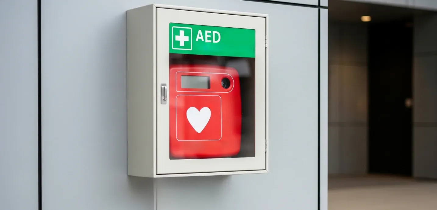
Hands-Only CPR and Defibrillation
Push hard and fast in the centre of the chest, aiming for 100 to 120 compressions per minute and pressing down by around 5 to 6 centimetres. Do not stop until the ambulance crew arrives and takes over. If a defibrillator (AED) is available nearby, send someone to retrieve it and use it as soon as possible. AEDs give voice instructions and are designed to be used by untrained bystanders. For a full guide to hands-only CPR technique and how to use a defibrillator, see our dedicated post: If Someone Collapses: What to Do Before the Ambulance Arrives.
Heart Attacks in the Workplace
Heart attacks do not observe working hours. The workplace is a setting where cardiac events occur, and where the presence of a trained first aider and an accessible defibrillator can mean the difference between survival and death.
Under the Health and Safety at Work Act 1974 and associated regulations, employers are required to make adequate first aid provision for their employees.[31] The HSE's guidance recommends that employers conduct a first aid needs assessment that accounts for the number and nature of employees, the work environment, and the distance from emergency services. A first aider trained to recognise and respond to cardiac emergencies is a meaningful component of that provision.
Public Access Defibrillation (PAD) schemes have expanded significantly in the UK, with defibrillators now present in many workplaces, shopping centres, sports facilities, and transport hubs.[32] Their presence saves lives, but only if people know they are there and are willing to use them. A first aider who knows where the nearest AED is located and is comfortable operating it is a genuine asset to any workplace.
A defibrillator is only useful if people know where it is, feel able to use it, and have practised the decision-making that happens under pressure. First aid training turns equipment on the wall into a usable emergency response.
Chronic work-related stress is also a cardiovascular risk factor. Employers who take workplace wellbeing seriously, not as a corporate exercise but as a genuine risk management responsibility, are reducing the background incidence of heart disease in their workforce alongside preparing to respond when acute events occur.
Life After a Heart Attack
Surviving a heart attack is not the end of the story. The weeks and months that follow are a critical period in which the risk of a second event is elevated, and in which the lifestyle changes, medications, and rehabilitation that reduce that risk need to be established.
Most people who survive a heart attack in the UK are offered cardiac rehabilitation: a structured programme of exercise, education, and psychological support that has strong evidence for reducing mortality and improving quality of life.[33] Uptake remains lower than it should be, particularly among women and people from minority ethnic backgrounds.
Standard secondary prevention medications typically include antiplatelet agents (such as aspirin and clopidogrel), beta-blockers, ACE inhibitors, and statins. These are not optional extras: they are treatments with robust evidence bases that substantially reduce the risk of further events.
The psychological impact of a heart attack is frequently underestimated. Depression affects around 20% of heart attack survivors and is itself associated with worse cardiovascular outcomes.[34] Anxiety about physical exertion, changes in self-perception, and the adjustment of relationships and identity following a significant health event are all real and common. Psychological support, whether through cardiac rehabilitation programmes, GP referral, or peer support networks, is part of recovery, not a soft add-on.
Could You Respond in a Cardiac Emergency?
Knowing the theory of a heart attack is one thing. Being able to act confidently and correctly in the moment is another. First aid training builds the practical skills (compression technique, defibrillator use, bystander decision-making) that translate knowledge into effective response.
Constellation Training offers first aid courses accredited by FAIB, covering cardiac emergency response alongside a full range of first aid competencies. Whether you are an employer reviewing first aid provision, or a team wanting people to respond with more confidence, our courses are designed to produce capable, composed responders.
Find out more about our first aid training courses and upcoming dates on our courses page.
This post is for information and general guidance only. It does not replace professional medical advice, diagnosis, or treatment. In an emergency, call 999.
References
[1] British Heart Foundation. Heart statistics. Available at: https://www.bhf.org.uk/what-we-do/our-research/heart-statistics [Accessed May 2026].
[2] World Health Organization. Cardiovascular diseases (CVDs). Fact sheet. Available at: https://www.who.int/news-room/fact-sheets/detail/cardiovascular-diseases-(cvds) [Accessed May 2026].
[3] Allam AH et al. Atherosclerosis in ancient Egyptian mummies: the Horus study. JACC Cardiovasc Imaging. 2011;4(4):315–327.
[4] Harvey W. Exercitatio Anatomica de Motu Cordis et Sanguinis in Animalibus. Frankfurt: 1628.
[5] Heberden W. Some account of a disorder of the breast. Medical Transactions of the Royal College of Physicians. 1772;2:59–67.
[6] Herrick JB. Clinical features of sudden obstruction of the coronary arteries. JAMA. 1912;59(23):2015–2020.
[7] Oxford Latin Dictionary. Infarctus. Oxford University Press.
[8] Einthoven W. The string galvanometer and the human electrocardiogram. Nobel Lecture, 1924.
[9] Julian DG. Treatment of cardiac arrest in acute myocardial ischaemia and infarction. Lancet. 1961;2:840–844.
[10] Fibrinolytic Therapy Trialists' Collaborative Group. Indications for fibrinolytic therapy in suspected acute myocardial infarction. Lancet. 1994;343:311–322.
[11] Brodie BR et al. Door-to-balloon time and clinical outcomes in patients with ST-elevation myocardial infarction. JACC. 2006;47(11):2180–2186.
[12] Libby P et al. Atherosclerosis. Nature Reviews Disease Primers. 2019;5:56.
[13] Falk E. Pathogenesis of atherosclerosis. J Am Coll Cardiol. 2006;47(8 Suppl):C7–12.
[14] Jennings RB, Reimer KA. The cell biology of acute myocardial ischemia. Annu Rev Med. 1991;42:225–246.
[15] NICE Clinical Guideline CG95. Chest pain of recent onset: assessment and diagnosis. NICE, 2016.
[16] British Heart Foundation. Delay in seeking medical help for heart attack symptoms. BHF report, 2019.
[17] NHS England. Myocardial Ischaemia National Audit Project (MINAP). 2023 report.
[18] Mehta LS et al. Acute myocardial infarction in women: a scientific statement from the American Heart Association. Circulation. 2016;133(9):916–947.
[19] Alabas OA et al. Sex differences in treatments, relative survival, and excess mortality following acute myocardial infarction. J Am Heart Assoc. 2017;6(4):e004375.
[20] Saw J et al. Spontaneous coronary artery dissection: association with predisposing arteriopathies and precipitating stressors and cardiovascular outcomes. Circ Cardiovasc Interv. 2014;7(5):645–655.
[21] Gholap N et al. Ethnic differences in incidence of type 2 diabetes and coronary heart disease in England and Wales. BMC Public Health. 2011;11:760.
[22] Rathmann W, Giani G. Global prevalence of diabetes: estimates for the year 2000 and projections for 2030. Diabetes Care. 2004;27(10):2568–2569.
[23] NHS Digital. Health Survey for England 2019: cardiovascular disease, diabetes and related risk factors.
[24] NICE. Cardiovascular disease risk assessment and reduction, including lipid modification. NICE Guideline NG238, 2023.
[25] Fleg JL et al. Detection of silent myocardial ischemia in a healthy elderly population. Circulation. 1990;81(2):428–436.
[26] NHS. Benefits of stopping smoking. Available at: https://www.nhs.uk/better-health/quit-smoking [Accessed May 2026].
[27] NHS Digital. Health Survey for England 2019: hypertension.
[28] Emerging Risk Factors Collaboration. Diabetes mellitus, fasting blood glucose concentration, and risk of vascular disease. Lancet. 2010;375(9733):2215–2222.
[29] Kivimäki M, Kawachi I. Work as a risk factor for cardiovascular disease. Curr Cardiol Rep. 2015;17(9):74.
[30] NICE Guideline NG185. Acute coronary syndromes. NICE, 2020. Available at: https://www.nice.org.uk/guidance/ng185 [Accessed May 2026].
[31] Health and Safety Executive. First aid at work: the Health and Safety (First-Aid) Regulations 1981. Available at: https://www.hse.gov.uk/firstaid [Accessed May 2026].
[32] Resuscitation Council UK. Public Access Defibrillation guidance, 2025. Available at: https://www.resus.org.uk/professional-library/2025-resuscitation-guidelines [Accessed May 2026].
[33] NICE Guideline NG172. Cardiac rehabilitation. NICE, 2020. Available at: https://www.nice.org.uk/guidance/ng172 [Accessed May 2026].
[34] Thombs BD et al. Prevalence of depression in survivors of acute myocardial infarction. J Gen Intern Med. 2006;21(1):30–38.